Your Microscope Is Finally Seeing What Matters
Watching tiny biological processes up close is vital for science, but current tools often miss the most important details. A new open-source system lets researchers track moving samples with incredible precision, changing how we understand life.
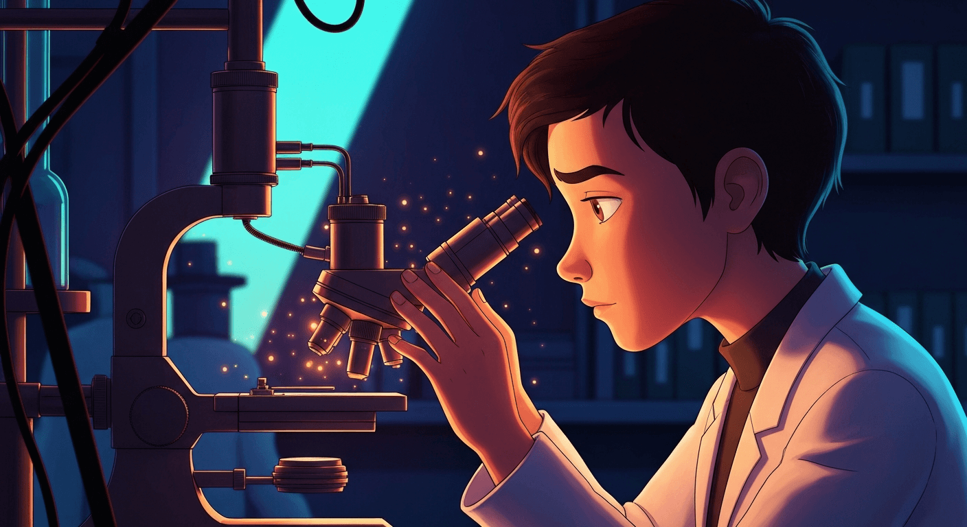
Have you ever tried to follow a fast-moving object in a crowd? It's tough, right? Even with today's sophisticated tools, scientists face a similar challenge when trying to observe living cells or tissues under a microscope. Imagine a tiny fish embryo developing: its cells aren't sitting still for a picture. They’re constantly shifting, moving, and interacting, making it incredibly difficult to capture those crucial, dynamic moments. This inability to reliably track a moving sample means researchers often miss key events, making their observations incomplete.
This isn't a simple "zoom in" problem. Traditional microscopy systems are good at taking static snapshots or fixed-point videos. But when the sample itself moves, the microscope objective – the lens array that focuses the light – can't always keep up. It's like trying to photograph a running dog with a camera fixed on a tripod; you might get blurry streaks or miss the dog entirely. Scientists have often resorted to tedious, manual adjustments, nudging the sample holder to keep their subject in view. This eats up valuable research time and can lead to inconsistent results.
That’s why a new tool called DySTrack is causing a quiet stir. It’s an open-source Python tool that lets commercial microscopes "see" and follow dynamic samples in real time. Think of it like giving your microscope a pair of intelligent eyes and a steady hand. Instead of just recording what's in front of it, it can now identify a specific moving cell or tissue and adjust its view to keep that target perfectly centered. This allows for continuous, high-resolution observation of living processes, offering insights previously hidden.
Giving Microscopes a Smart Brain
DySTrack works by essentially building a bridge between the physical microscope and powerful computer analysis. Most high-end microscopes come with their own software for setting up shots and collecting images. DySTrack doesn't replace that; instead, it plugs into it, taking the live images and feeding them into an external "brain"—a powerful image analysis pipeline running on a computer. This pipeline quickly processes the images, figures out where the target object has moved, and then sends instructions back to the microscope to re-center the view. This all happens in milliseconds, allowing for smooth, continuous tracking.
It’s a bit like a chef who loves their familiar kitchen appliances but wants to try a new, complex recipe that requires precise timing and ingredient tracking. Instead of buying all new appliances, they bring in a smart assistant who watches the cooking, gives real-time feedback, and adjusts temperatures or timings on the existing equipment. Researchers get to use their comfortable, familiar microscope controls, while gaining the muscle of advanced, custom image analysis . This modular approach makes it easier for labs to adopt without a complete overhaul of their existing setup.
Watching Life Unfold, Not Just Freeze-Frames
What does this mean for understanding life itself? Take the development of an embryo. Scientists can now track individual cell groups, like the lateral line primordium in a zebrafish or Hensen's node in a chick, as they migrate and form complex structures. This is like finally being able to watch an entire symphony from start to finish, rather than just hearing isolated notes. You can observe the precise dance of cells forming tissues, organs taking shape, and how external factors might influence these delicate processes over time. Before, such long-term, high-resolution tracking was incredibly challenging and often impossible without manual intervention.
One surprising fact about this kind of "smart microscopy" is how much data it generates. A single, continuous tracking experiment can produce terabytes of images, far more than traditional methods. Processing this massive amount of visual information is where the external Python analysis steps in, handling the computational heavy lifting to extract meaningful patterns. This capability also makes it much easier to quantify cellular movements and interactions, giving scientists hard numbers instead of just qualitative observations.
What Comes Next for Smart Microscopy
While DySTrack makes smart microscopy more accessible, it's still an area that needs specialists to set up the initial analysis pipelines. However, its open-source nature means that researchers worldwide can contribute to and improve it, creating more ready-to-use "recipes" for different biological questions. This kind of collaborative development is crucial for spreading the technology. You can expect to see wider adoption in academic labs focused on developmental biology, neuroscience, and disease modeling in the next 5-10 years.
This isn't just about fancier microscopes; it's about fundamentally changing how we approach biological discovery. By enabling automated, precise observation of living systems, DySTrack allows scientists to ask deeper questions about how life works, how diseases progress, and how organisms develop. It helps reveal the intricate choreography of cells, unlocking secrets that were previously invisible because our tools couldn’t keep pace with life’s constant motion. Your understanding of biology is about to get a whole lot clearer. .

Key Takeaways
- Traditional microscopes struggle to track moving biological samples, leading to missed observations of key cellular processes.
- DySTrack offers an open-source solution that links commercial microscopes with powerful external image analysis, enabling real-time, automated tracking.
- This innovation allows scientists to observe dynamic biological events, such as embryo development, with unprecedented precision, leading to deeper insights into life's mechanisms.
Frequently Asked Questions
What is smart microscopy? Smart microscopy uses computer analysis to interpret images in real-time and automatically adjust the microscope's settings, like focus or position, to follow moving samples. It makes observation more precise and automated.
How does DySTrack help researchers? DySTrack connects commercial microscopes with flexible image analysis software, allowing scientists to track moving biological samples like developing embryos. This eliminates manual adjustments and reveals dynamic cellular processes more clearly.
Why is tracking moving samples important? Living biological samples are constantly changing. Tracking their movement continuously helps scientists understand dynamic processes like cell migration, tissue development, and disease progression, providing a more complete picture of life.
Editorial note: The scientific findings presented in this article are sourced exclusively from published research papers, peer-reviewed studies, certified inventions, and registered patent filings. Images generated by AI.
Stay ahead of the curve
The science that shapes tomorrow — in your inbox every week
The scientific findings presented in our articles are sourced from published research papers, peer-reviewed studies, certified inventions, and registered patent filings. Subscribe for focused weekly coverage, hands-on explainers, and practical insights that help you stay curious — no jargon, no noise.
By subscribing, you agree to receive newsletter and marketing emails, and accept our Terms of Use and Privacy Policy. You can unsubscribe anytime.
AI Ethics, Algorithmic Bias & Responsible Computing
Technology ethicist and journalist covering the human consequences of the decisions embedded in algorithms and AI systems.
View full profile →