AI Can Now Spot Cancer Cells Doctors Miss
Tiny, early-stage cancer cells often slip past standard screening. New AI models are catching what human eyes miss, cutting diagnosis time and sharpening treatment precision.

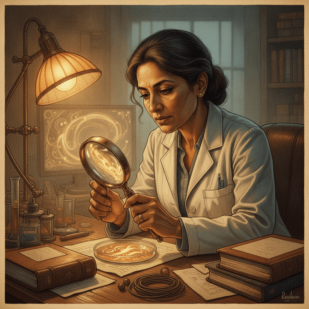
Doctors might soon be able to find hidden cancer cells in your body with incredible speed and accuracy, thanks to advanced AI that acts like a super-sharp second pair of eyes. Think of this AI as a highly trained detective, capable of sifting through vast amounts of information – in this case, microscopic images of your tissues – to pinpoint clues that even experienced human eyes might overlook. This isn't science fiction; it’s being developed right now.
Right now, most digital pathology tools are good at looking at small sections of a tissue sample, like zoomed-in snapshots. However, they often struggle with the entire "whole-slide image," which is like trying to understand a whole city by only looking at individual street corners. But new models called GigaPath-Flash and GigaTIME-Flash are changing this, bringing the power of "foundation models"—which are large AI systems trained on massive amounts of data, like a language model learning from millions of books—to the complex world of cancer diagnosis.
These models are like expert cartographers, able to analyze the entire map of your tissue sample, not just a few districts. They've been trained on real-world medical data, allowing them to learn the intricate patterns of disease. One surprising fact: the GigaPath-Flash model achieves 97% of the performance of a much larger, more expensive predecessor, but uses 50 times less computing power, making it far more practical for widespread use in hospitals.
So, how do these systems actually work? At their core, they use what's called a Vision Transformer (ViT-S) as a "tile encoder." Imagine you have a giant mosaic, and this encoder is a specialist in understanding each tiny tile, piece by piece. After understanding the individual tiles, another part, called a LongNet slide encoder, then takes all that tile information and pieces together the entire mosaic, understanding the big picture of the whole tissue slide. This two-step process allows the AI to see both the fine details and the overall context, crucial for spotting elusive signs of disease.
This isn't just about spotting cancer; it's about understanding its environment. GigaTIME-Flash, a sibling model, goes a step further by predicting the "tumor immune microenvironment" directly from standard tissue images. Think of this as getting a detailed weather report for the tiny ecosystem surrounding a tumor—how the immune cells are interacting with the cancer. This is vital because the immune environment plays a huge role in how a tumor behaves and how it responds to treatments, including your body can finally target sickness. Knowing this helps doctors tailor therapies more precisely.
Currently, these models are in the preprint stage, meaning they've been shared with the scientific community but haven't yet undergone the full peer-review process that leads to publication in a journal. However, the researchers behind GigaPath-Flash and GigaTIME-Flash have made all their models and training weights openly available with an Apache-2.0 license. This is a big deal because it means other researchers and institutions can pick up these tools and start using them, testing them, and building upon them without restrictive access or fees.
This open-source approach will accelerate development and adoption. What’s holding it back? Large-scale clinical validation in diverse hospital settings is the next big step. This involves testing the AI on thousands, even millions, of patient samples to ensure it performs consistently across different patient populations and equipment. It also requires integrating these complex AI tools into existing hospital workflows, which can be a significant logistical challenge, often requiring new smarter AI for doctors.
If clinical trials go well and regulatory bodies approve these methods, you could see AI like this assisting your pathologist in cancer diagnoses within the next 5-7 years. This means faster, more accurate diagnoses, especially for tiny, early-stage cancers that are harder to detect. Imagine getting a diagnosis not just earlier, but with a deeper understanding of your tumor's specific biology, paving the way for more personalized and effective treatment plans. It means a future where your doctor has an incredibly powerful ally in the fight against disease, quietly working behind the scenes to give you the best possible outcome.
Understanding How AI "Sees" Disease
AI in pathology uses sophisticated algorithms to analyze digital images of tissue. It breaks down complex visual data into patterns and features, much like a human eye recognizes shapes and colors, but with far greater precision and speed. This helps identify subtle abnormalities.
Specifically, AI is trained on enormous datasets of correctly diagnosed cancer and healthy tissue samples. It learns to differentiate between various cell types, tissue structures, and even the subtle indicators of disease progression. This training allows the AI to develop an "eye" for what to look for, surpassing what even the most diligent human can process in the same timeframe. This includes identifying specific molecular markers that might indicate tumors hiding from treatment.
The Power of Open-Source Medical AI
Making medical AI models open-source dramatically speeds up their development and integration into healthcare. When models like GigaPath-Flash and GigaTIME-Flash are freely available, researchers worldwide can immediately begin using them, validating them with their own datasets, and contributing improvements. This collaborative approach means that the technology can evolve much faster than if it were held under proprietary licenses by a single company. It democratizes access to advanced tools, fostering innovation globally.

Key Takeaways
- New AI models, GigaPath-Flash and GigaTIME-Flash, efficiently analyze entire tissue slides to detect hidden cancer cells.
- These AI systems can predict the tumor's immune environment, offering crucial insights for personalized cancer treatment plans.
- The open-source release of these models will significantly accelerate their development, validation, and adoption in clinical settings globally.
Frequently Asked Questions
What is a foundation model in pathology? A foundation model in pathology is a large AI system trained on vast amounts of medical imaging data. It learns to recognize complex patterns in tissue samples, enabling it to assist in tasks like cancer diagnosis and understanding disease characteristics.
How do GigaPath-Flash and GigaTIME-Flash improve diagnosis? These models analyze entire digital tissue slides, not just small sections, allowing them to spot hidden cancer cells and understand the tumor's environment more comprehensively. This leads to faster, more accurate detection and better treatment planning.
When could this AI be used in hospitals? After further clinical validation and regulatory approvals, which could take 5-7 years, this type of AI could become a standard tool in pathology labs, assisting doctors with diagnostics and treatment decisions.
Editorial note: The scientific findings presented in this article are sourced exclusively from published research papers, peer-reviewed studies, certified inventions, and registered patent filings. Images generated by AI.
Stay ahead of the curve
The science that shapes tomorrow — in your inbox every week
The scientific findings presented in our articles are sourced from published research papers, peer-reviewed studies, certified inventions, and registered patent filings. Subscribe for focused weekly coverage, hands-on explainers, and practical insights that help you stay curious — no jargon, no noise.
By subscribing, you agree to receive newsletter and marketing emails, and accept our Terms of Use and Privacy Policy. You can unsubscribe anytime.
AI in Healthcare, Biomedical Computing & Drug Discovery Algorithms
Computational biologist and science journalist covering the remarkable collision of artificial intelligence with medical research.
View full profile →