Your X-Rays May Soon Not Need Radiation
Did you know that children's hospital visits for broken bones expose them to radiation that builds up over a lifetime? A new AI-powered imaging method promises to show bone injuries without a single X-ray.
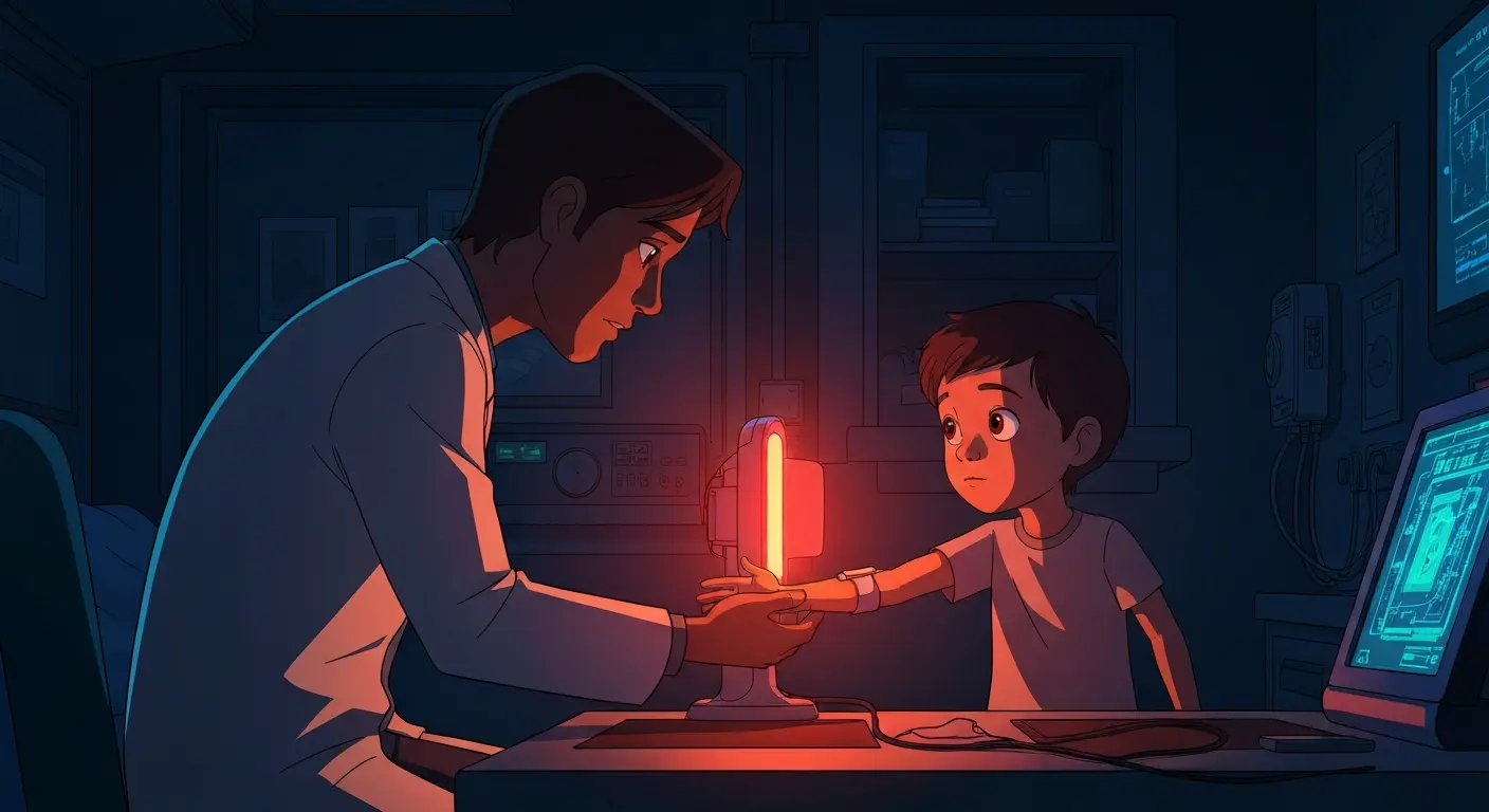
You might soon see inside your body without the ionizing radiation of traditional X-rays. For years, medical imaging has relied on X-rays to peer through skin and muscle to see bones, but this method carries a small, cumulative risk, especially for children. What if a new technique could show bone injuries as clearly as an X-ray, but simply by reading the heat and light reflecting off your skin?
This isn't science fiction; it's a future that's much closer than you think, thanks to a clever pairing of infrared light and artificial intelligence. Researchers are developing a way to map the internal structures of your body, like your bones, using different types of infrared light. Imagine a kind of super-sensitive thermal camera that can see not just surface heat, but also how light behaves deep within your tissues. The goal is to build detailed "synthetic radiographs" – digital images that look just like X-rays, but without using any harmful radiation.
Sensing Hidden Signals with Infrared Light
The core idea is to use different "colors" of infrared light, which are like invisible light rays on the electromagnetic spectrum that we perceive as heat. These different infrared wavelengths can penetrate tissues at varying depths. Some go just under the skin, while others can reach much deeper, almost like tiny probes. Think of it like a chef adding different spices to a dish: each one brings out a distinct flavor or characteristic. Similarly, each infrared wavelength brings back unique data about your body's internal composition.
For instance, near-infrared (NIR) light, which is close to the visible light spectrum, can pass through skin and even bone, especially in smaller structures like a child's forearm. Scientists have already used similar light for things like monitoring brain activity, showing how well it can travel through the body. By bouncing these different infrared lights off your body and capturing the reflections, doctors can gather a wealth of data about what's happening underneath the surface. This technique could be particularly valuable for children, where repeated X-ray exposure raises concerns about long-term health risks like leukemia. One surprising fact: over 18% of all pediatric emergency room visits are for musculoskeletal trauma, making this a common concern.

How AI Turns Invisible Light Into Visible Bones
Collecting all that infrared data is just the first step. The real magic happens when artificial intelligence, specifically a type of AI called a deep-learning cross-modal translation network, gets involved. These networks are like highly skilled translators. They take the raw, complex infrared data – which looks nothing like a typical X-ray – and "translate" it into a clear, clinically usable image that resembles an X-ray. They learn to identify patterns in the infrared signals that correspond to bone, muscle, and other tissues.
Think of it this way: imagine you have a very detailed, blurry photograph, and an AI that's been trained on millions of sharp photographs. The AI can then use its knowledge to "unblur" your picture and even fill in missing details, making it look incredibly crisp. In this case, the AI is essentially "unblurring" the hidden infrared signals to create a sharp image of your bones. Researchers are using advanced AI models, like those named Pix2Pix and CycleGAN, to perform this complex image-to-image translation. This means your doctor could potentially see a broken bone on a screen, generated solely from a harmless light scan, without ever taking an X-ray.
The Path to Radiation-Free Imaging
Currently, this technology is in the research and development phase, with teams focusing on building the essential datasets needed to train these powerful AIs. To truly work, the AI needs to see countless examples of paired infrared scans and traditional X-rays from real patients. This teaches it exactly how to create a "synthetic" X-ray from the infrared data. The ultimate goal is to make these devices compact and portable, potentially allowing for quick, safe triage of injuries right in a doctor's office or even a sports field.
What’s holding it back? One major hurdle is gathering enough high-quality, matched datasets – meaning the same patient gets both an infrared scan and a traditional X-ray, allowing the AI to learn the correlation. Another challenge involves regulatory approvals for AI as a medical device, and ensuring the models work reliably across different body types and skin tones. However, if these challenges are met, you could see such radiation-free imaging systems in pediatric clinics and emergency rooms within the next 10-15 years. This could fundamentally change how we approach diagnosing bone injuries, particularly for the youngest patients, offering a safer alternative to conventional methods.
Key Takeaways
- New imaging combines harmless infrared light with AI to create "synthetic X-rays" of bones.
- This approach completely avoids the radiation risks associated with traditional X-rays, especially beneficial for children.
- The technology is still in development, primarily focused on building large datasets and securing regulatory approval, with an estimated availability of 10-15 years.
Frequently Asked Questions
What is infrared imaging for bones? It's a medical technique using different types of invisible infrared light to scan the body. The light penetrates tissues and its reflections are read by AI to create detailed images of bones without X-ray radiation.
How does AI help create bone images from infrared? Special AI models are trained on countless examples of infrared scans and matching X-rays. They learn to translate the invisible infrared data into clear, visible images that look just like traditional X-rays.
When might this technology be available? While in development, the technology faces challenges like data collection and regulatory approval. If progress continues steadily, radiation-free bone imaging could be a reality in clinics and hospitals within the next 10-15 years.
Editorial note: The scientific findings presented in this article are sourced exclusively from published research papers, peer-reviewed studies, certified inventions, and registered patent filings. Images generated by AI.
Stay ahead of the curve
The science that shapes tomorrow — in your inbox every week
The scientific findings presented in our articles are sourced from published research papers, peer-reviewed studies, certified inventions, and registered patent filings. Subscribe for focused weekly coverage, hands-on explainers, and practical insights that help you stay curious — no jargon, no noise.
By subscribing, you agree to receive newsletter and marketing emails, and accept our Terms of Use and Privacy Policy. You can unsubscribe anytime.
AI in Healthcare, Biomedical Computing & Drug Discovery Algorithms
Computational biologist and science journalist covering the remarkable collision of artificial intelligence with medical research.
View full profile →