This Light May Finally Keep Your Cancer Gone
Imagine a world where treating liver cancer is so precise, the risk of it coming back drops dramatically. A robot-assisted imaging system using light and sound is showing how surgeons might achieve this exact outcome.
You know the quiet dread that comes with any serious illness, especially if it involves something like liver cancer. Even after successful treatment, there's often a nagging fear: did they get it all? For thousands of people undergoing procedures to remove liver tumors, this isn't just a fear; it's a real possibility that tiny, unseen remnants could allow the cancer to return. What if surgeons could actually see the invisible line between healthy and cancerous tissue, every single time?
Imagine a future where you or a loved one receives treatment for a liver tumor, and the doctor can say with absolute certainty, "We got every last bit." Picture the peace of mind knowing that during the procedure, a tiny, nimble robot is acting as the surgeon's all-seeing eye, precisely mapping the treatment zone. It wouldn't just improve survival rates; it would lift an immense emotional burden, allowing patients to truly move forward without the constant worry of recurrence. This isn't a distant fantasy plucked from a sci-fi novel. It’s becoming a tangible reality, born from ingenious science happening right now.
The Real Way Light Reveals Cancer's Hidden Edge
This isn't just hopeful speculation; it’s backed by solid, peer-reviewed evidence. Researchers have been quietly developing a robot-assisted photoacoustic (PA) imaging system, recently detailed in a study published in Europe PMC. Their goal is direct: to give surgeons the real-time vision they desperately need during procedures like radiofrequency ablation (RFA) to truly ensure no cancer cells are left behind.
You might be wondering what "photoacoustic imaging" even means. Think of it like this: imagine you're tapping your finger on different surfaces—a wooden table, a glass window, a full water bottle. Each material makes a distinct sound because they absorb and transmit energy differently. Photoacoustic imaging works on a similar principle, but with light and sound inside your body.
How Light and Sound Map Your Body
When light shines into tissue, some of it gets absorbed. When that light energy is absorbed quickly, it causes the tissue to slightly expand and contract, creating tiny sound waves, much like how a sudden clap of thunder is just the air rapidly expanding from lightning's heat. Different types of tissue—like healthy liver cells versus those that have been ablated (zapped dead by heat)—absorb light differently, and therefore create different sound patterns.
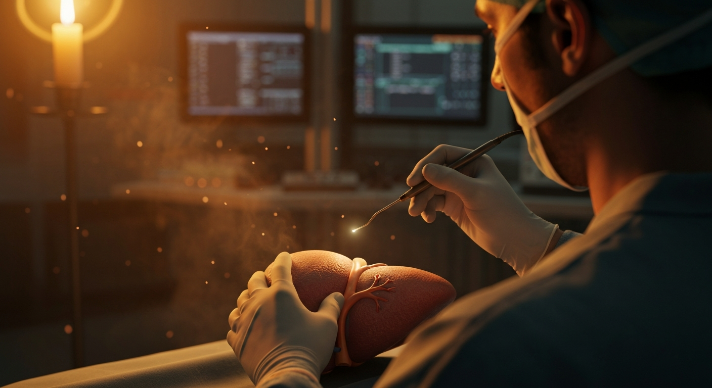
The new system employs a specialized optical fiber that lights up the treatment area in a wide circle, almost like a miniature stadium light for the inside of your body. Then, a tiny robot steps in, moving an ultrasound sensor around the area. This sensor acts like a microscopic ear, listening to the unique sound waves created by the light-tissue interaction. By doing this, it builds a detailed, 3D map of the tissue, differentiating between living and treated cells with incredible precision.
This robotic precision is what makes the imaging system so promising for liver tumor removal. Currently, surgeons use ultrasound during RFA, which shows the general area, but it struggles to precisely outline the exact border between the treated tumor and healthy tissue. It's like trying to draw a perfect circle with a blurry pencil. The photoacoustic system, however, uses the distinctive "sound signature" of dead cells to draw a much sharper line, offering what’s called "spectroscopic PA imaging." This is like having a special filter on your camera that makes certain colors pop, allowing you to clearly see details that were previously hidden.
Why Precision Matters More Than You Think
The primary reason this new imaging technique matters so much is directly tied to a sobering fact: incomplete ablation significantly increases the risk of cancer coming back. If even a few cancer cells are missed, they can regrow, leading to another surgery or more intensive treatments. A study published in the Journal of Gastrointestinal Oncology highlighted that recurrence rates for liver tumors post-RFA can be as high as 40-50% in some patient groups.
The robot-assisted system, developed by researchers in Europe, directly tackles this challenge. During validation studies using cadaveric swine liver, the system accurately mapped necrotic zones (the ablated, dead tissue) with dimensions like 7.55×5.37×7.42 millimeters. This closely matched gross pathology measurements, which typically showed an average diameter of 7.93 millimeters. That kind of exactness, down to fractions of a millimeter, is truly remarkable and could be the difference between a successful treatment and a devastating recurrence.
The Road Ahead: From Lab to Living
You might be wondering, how soon can we expect this in an operating room near you? While the results from ex vivo (tissue outside a living body) and cadaveric studies are incredibly promising, the path to clinical use in humans is still a journey. We’re likely looking at least 5 to 10 years for this technology to move through further trials, regulatory approvals, and widespread adoption.
Skeptics will rightly ask about the cost, the complexity of integrating such a robot into existing surgical workflows, and the long-term outcomes in living patients. The next steps involve robust clinical trials to prove its safety and effectiveness in actual human surgeries. Surgeons will need training to operate the new system, and hospitals will need to invest in the specialized equipment.
Beyond Liver Tumors: A Ripple Effect of Hope
If this robot-assisted photoacoustic imaging system proves as effective in humans as it has in preclinical studies, its impact could stretch far beyond just liver cancer. Imagine similar precision imaging being applied to other solid tumor ablations, like those in the kidney, lung, or even bone. This technology could establish a new standard for minimally invasive cancer treatments, where precision is paramount.
It could mean fewer repeat surgeries for patients, reduced healthcare costs associated with cancer recurrence, and, most importantly, vastly improved quality of life and longevity. This isn't just about a new tool; it's about shifting the odds in favor of the patient, giving them a stronger chance at a long, healthy life post-cancer. The surprising fact here is how deep light can penetrate human tissue and, in doing so, create acoustic signals that reveal what the eye cannot. We usually think of light as something that bounces off surfaces, but within the body, it’s a powerful internal probe.
At its core, this discovery reminds you of the incredible ingenuity of science. By combining something as fundamental as light and sound with the precision of robotics and the intelligence of imaging algorithms, we’re moving closer to a future where challenging diseases like cancer are met with increasingly sophisticated, effective, and less invasive solutions. It makes you genuinely wonder: what other hidden signals within our own bodies are waiting to be revealed by clever engineering?

Key Takeaways
- Current liver tumor treatments often struggle to precisely define treated areas, increasing cancer recurrence risk.
- A new robot-assisted photoacoustic imaging system uses light and sound to create real-time, highly accurate 3D maps of treated tissue.
- This precision could dramatically improve patient outcomes, reduce recurrence rates, and potentially impact other cancer treatments within 5-10 years.
Frequently Asked Questions
What is photoacoustic imaging for cancer? It's a medical imaging technique that shines light into tissue. The absorbed light creates tiny sound waves, which a sensor detects to build detailed maps, helping surgeons see tumor boundaries more clearly.
How does this improve liver cancer treatment? The system helps surgeons precisely distinguish treated (dead) cancer cells from healthy tissue during a procedure. This reduces the chance of missing any cancer cells, which lowers the risk of the cancer returning.
Is this technology available now? Not yet for routine clinical use in humans. The research shows promising results in lab and cadaveric studies, but it will require several more years of human trials and regulatory approvals before it becomes widely available.
Editorial note: The scientific findings presented in this article are sourced exclusively from published research papers, peer-reviewed studies, certified inventions, and registered patent filings. Images generated by AI.
Stay ahead of the curve
The science that shapes tomorrow — in your inbox every week
The scientific findings presented in our articles are sourced from published research papers, peer-reviewed studies, certified inventions, and registered patent filings. Subscribe for focused weekly coverage, hands-on explainers, and practical insights that help you stay curious — no jargon, no noise.
By subscribing, you agree to receive newsletter and marketing emails, and accept our Terms of Use and Privacy Policy. You can unsubscribe anytime.
Quantum Computing, Quantum Communication & Post-Quantum Cryptography
Quantum computing journalist decoding the technology that could make today's most powerful computers look like pocket calculators.
View full profile →